Agarose Gel Electrophoresis - Principle, Parts, Protocol, Applications, Advantages, Disadvantages
Agarose gel electrophoresis is the most effective way of separating DNA fragments of varying sizes ranging from 100 bp to 25 kb. Agarose is isolated from the seaweed genera Gelidium and Gracilaria, and consists of repeated agarobiose (L- and D-galactose) subunits. During gelation, agarose polymers associate non-covalently and form a network of bundles whose pore sizes determine a gel's molecular sieving properties. The use of agarose gel electrophoresis revolutionized the separation of DNA. Prior to the adoption of agarose gels, DNA was primarily separated using sucrose density gradient centrifugation, which only provided an approximation of size. To separate DNA using agarose gel electrophoresis, the DNA is loaded into pre-cast wells in the gel and a current applied. The phosphate backbone of the DNA (and RNA) molecule is negatively charged, therefore when placed in an electric field, DNA fragments will migrate to the positively charged anode. Because DNA has a uniform mass/charge ratio, DNA molecules are separated by size within an agarose gel in a pattern such that the distance traveled is inversely proportional to the log of its molecular weight. The leading model for DNA movement through an agarose gel is "biased reptation", whereby the leading edge moves forward and pulls the rest of the molecule along. The rate of migration of a DNA molecule through a gel is determined by the following: 1) size of DNA molecule; 2) agarose concentration; 3) DNA conformation; 4) voltage applied, 5) presence of ethidium bromide, 6) type of agarose and 7) electrophoresis buffer. After separation, the DNA molecules can be visualized under uv light after staining with an appropriate dye. By following this protocol, students should be able to: 1. Understand the mechanism by which DNA fragments are separated within a gel matrix 2. Understand how conformation of the DNA molecule will determine its mobility through a gel matrix 3. Identify an agarose solution of appropriate concentration for their needs 4. Prepare an agarose gel for electrophoresis of DNA samples 5. Set up the gel electrophoresis apparatus and power supply 6. Select an appropriate voltage for the separation of DNA fragments 7. Understand the mechanism by which ethidium bromide allows for the visualization of DNA bands 8. Determine the sizes of separated DNA fragments
What is Agarose Gel Electrophoresis?
- Agarose gel electrophoresis is a method of gel electrophoresis used in biochemistry, molecular biology, genetics, and clinical chemistry to separate a mixed population of macromolecules such as DNA , RNA or proteins in a matrix of agarose.
- Agarose is a natural linear polymer extracted from seaweed that forms a gel matrix by hydrogen-bonding when heated in a buffer and allowed to cool.
- They are the most popular medium for the separation of moderate and large-sized nucleic acids and have a wide range of separation.
Principle of Agarose Gel Electrophoresis
- Gel electrophoresis separates DNA fragments by size in a solid support medium such as an agarose gel. Sample (DNA) are pipetted into the sample wells, followed by the application of an electric current which causes the negatively-charged DNA to migrate (electrophorese) towards the anodal, positive (+ve) end. The rate of migration is proportional to size: smaller fragments move more quickly and wind up at the bottom of the gel.
- DNA is visualized by including in the gel an intercalating dye, ethidium bromide. DNA fragments take up the dye as they migrate through the gel. Illumination with ultraviolet light causes the intercalated dye to fluoresce.
- The larger fragments fluoresce more intensely. Although each of the fragments of a single class of molecule is present in equimolar proportions, the smaller fragments include less mass of DNA, take up less dye, and therefore fluoresce less intensely. A “ladder” set of DNA fragments of known size can be run simultaneously and used to estimate the sizes of the other unknown fragments.
Requirements/ Instrumentation of Agarose Gel Electrophoresis
The equipment and supplies necessary for conducting agarose gel electrophoresis are relatively simple and include:
- An electrophoresis chamber and power supply
- Gel casting trays, which are available in a variety of sizes and composed of UVtransparent plastic. The open ends of the trays are closed with tape while the gel is being cast, then removed prior to electrophoresis.
- Sample combs, around which molten medium is poured to form sample wells in the gel.
- Electrophoresis buffer, usually Tris-acetate-EDTA (TAE) or Tris-borate-EDTA (TBE).
- Loading buffer, which contains something dense (e.g. glycerol) to allow the sample to “fall” into the sample wells, and one or two tracking dyes, which migrate in the gel and allow visual monitoring or how far the electrophoresis has proceeded.
- Staining: DNA molecules are easily visualized under an ultraviolet lamp when electrphoresed in the presence of the extrinsic fluor ethidium bromide. Alternatively, nucleic acids can be stained after electrophoretic separation by soaking the gel in a solution of ethidium bromide. When intercalated into doublestranded DNA, fluorescence of this molecule increases greatly. It is also possible to detect DNA with the extrinsic fluor 1-anilino 8-naphthalene sulphonate.
- Transilluminator (an ultraviolet light box), which is used to visualize stained DNA in gels.
Steps Involved in Agarose Gel Electrophoresis
1. To prepare gel, agarose powder is mixed with
electrophoresis buffer to the desired concentration, and heated in a microwave
oven to melt it.
The concentration of Agarose Gel
- The percentage of agarose used depends on the size of fragments to be resolved.
- The concentration of agarose is referred to as a percentage of agarose to volume of buffer (w/v), and agarose gels are normally in the range of 0.2% to 3%.
- The lower the concentration of agarose, the faster the DNA fragments migrate.
- In general, if the aim is to separate large DNA fragments, a low concentration of agarose should be used, and if the aim is to separate small DNA fragments, a high concentration of agarose is recommended.
2. Ethidium bromide is added to the gel (final concentration
0.5 ug/ml) to facilitate visualization of DNA after electrophoresis.
3. After cooling the solution to about 60oC, it is poured
into a casting tray containing a sample comb and allowed to solidify at room
temperature.
4. After the gel has solidified, the comb is removed, taking
care not to rip the bottom of the wells.
5. The gel, still in plastic tray, is inserted horizontally
into the electrophoresis chamber and is covered with buffer.
6. Samples containing DNA mixed with loading buffer are then
pipetted into the sample wells, the lid and power leads are placed on the
apparatus, and a current is applied.
7. The current flow can be confirmed by observing bubbles
coming off the electrodes.
8. DNA will migrate towards the positive electrode, which is
usually colored red, in view of its negative charge.
9. The distance DNA has migrated in the gel can be judged by
visually monitoring migration of the tracking dyes like bromophenol blue and
xylene cyanol dyes.
Protocol of Agarose Gel Electrophoresis
1. Preparation of the Gel
- Weigh out the appropriate mass of agarose into an Erlenmeyer flask. Agarose gels are prepared using a w/v percentage solution. The concentration of agarose in a gel will depend on the sizes of the DNA fragments to be separated, with most gels ranging between 0.5%-2%. The volume of the buffer should not be greater than 1/3 of the capacity of the flask.
- Add running buffer to the agarose-containing flask. Swirl to mix. The most common gel running buffers are TAE (40 mM Tris-acetate, 1 mM EDTA) and TBE (45 mM Tris-borate, 1 mM EDTA).
- Melt the agarose/buffer mixture. This is most commonly done by heating in a microwave, but can also be done over a Bunsen flame. At 30 s intervals, remove the flask and swirl the contents to mix well. Repeat until the agarose has completely dissolved.
- Add ethidium bromide (EtBr) to a concentration of 0.5 μg/ml. Alternatively, the gel may also be stained after electrophoresis in running buffer containing 0.5 μg/ml EtBr for 15-30 min, followed by destaining in running buffer for an equal length of time.
Note: EtBr is a suspected carcinogen and must be properly disposed of per institution regulations. Gloves should always be worn when handling gels containing EtBr. Alternative dyes for the staining of DNA are available; however EtBr remains the most popular one due to its sensitivity and cost.
- Allow the agarose to cool either on the benchtop or by incubation in a 65 °C water bath. Failure to do so will warp the gel tray.
- Place the gel tray into the casting apparatus. Alternatively, one may also tape the open edges of a gel tray to create a mold. Place an appropriate comb into the gel mold to create the wells.
- Pour the molten agarose into the gel mold. Allow the agarose to set at room temperature. Remove the comb and place the gel in the gel box. Alternatively, the gel can also be wrapped in plastic wrap and stored at 4 °C until use
2. Setting up of Gel Apparatus and Separation of DNA
Fragments
- Add loading dye to the DNA samples to be separated. Gel loading dye is typically made at 6X concentration (0.25% bromphenol blue, 0.25% xylene cyanol, 30% glycerol). Loading dye helps to track how far your DNA sample has traveled, and also allows the sample to sink into the gel.
- Program the power supply to desired voltage (1-5V/cm between electrodes).
- Add enough running buffer to cover the surface of the gel. It is important to use the same running buffer as the one used to prepare the gel.
- Attach the leads of the gel box to the power supply. Turn on the power supply and verify that both gel box and power supply are working.
- Remove the lid. Slowly and carefully load the DNA sample(s) into the gel. An appropriate DNA size marker should always be loaded along with experimental samples.
- Replace the lid to the gel box. The cathode (black leads) should be closer the wells than the anode (red leads). Double check that the electrodes are plugged into the correct slots in the power supply.
- Turn on the power. Run the gel until the dye has migrated to an appropriate distance.
3. Observing Separated DNA fragments
- When electrophoresis has completed, turn off the power supply and remove the lid of the gel box.
- Remove gel from the gel box. Drain off excess buffer from the surface of the gel. Place the gel tray on paper towels to absorb any extra running buffer.
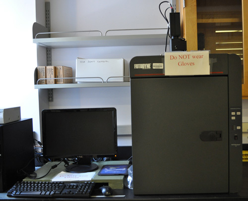
- Remove the gel from the gel tray and expose the gel to uv light. This is most commonly done using a gel documentation system. DNA bands should show up as orange fluorescent bands. Take a picture of the gel
- Properly dispose of the gel and running buffer per institution regulations.
4. Representative Results
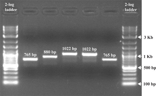
Represents a typical result after agarose gel
electrophoresis of PCR products. After separation, the resulting DNA fragments
are visible as clearly defined bands. The DNA standard or ladder should be
separated to a degree that allows for the useful determination of the sizes of
sample bands. In the example shown, DNA fragments of 765 bp, 880 bp and 1022 bp
are separated on a 1.5% agarose gel along with a 2-log DNA ladder.
Applications of Agarose Gel Electrophoresis
Agarose gel electrophoresis is a routinely used method for separating proteins, DNA or RNA.
- Estimation of the size of DNA molecules
- Analysis of PCR products, e.g. in molecular genetic diagnosis or genetic fingerprinting
- Separation of restricted genomic DNA prior to Southern analysis, or of RNA prior to Northern analysis.
- The agarose gel electrophoresis is widely employed to estimate the size of DNA fragments after digesting with restriction enzymes, e.g. in restriction mapping of cloned DNA.
- Agarose gel electrophoresis is commonly used to resolve circular DNA with different supercoiling topology, and to resolve fragments that differ due to DNA synthesis.
- In addition to providing an excellent medium for fragment size analyses, agarose gels allow purification of DNA fragments. Since purification of DNA fragments size separated in an agarose gel is necessary for a number molecular techniques such as cloning, it is vital to be able to purify fragments of interest from the gel.
Advantages of Agarose Gel Electrophoresis
- For most applications, only a single-component agarose is needed and no polymerization catalysts are required. Therefore, agarose gels are simple and rapid to prepare.
- The gel is easily poured, does not denature the samples.
- The samples can also be recovered.
Disadvantages of Agarose Gel Electrophoresis
- Gels can melt during electrophoresis.
- The buffer can become exhausted.
- Different forms of genetic material may run in unpredictable forms.
References
https://pdfs.semanticscholar.org/36cf/d4ada922c44d233b6ebfa2af2c956c92e4ec.pdf
https://www.mun.ca/biology/scarr/Gel_Electrophoresis.html
https://www.wou.edu/las/physci/ch462/Gel%20Electrophoresis.pdf


No comments