Lac Operon – Overview
What is lac Operon?
- The lactose operon (also known as the lac operon) is a set of genes that are specific for uptake and metabolism of lactose and is found in E. coli and other bacteria.
- Lac operon is a set of genes with a single promoter that codes for lactose transport and metabolism in E. coli and other bacteria.
- The lactose operon (lac operon) is an operon that E. coli and many other enteric bacteria need to move lactose around and break it down.
- Even though glucose is the preferred carbon source for most bacteria, the lac operon makes it possible for beta-galactosidase to break down lactose when glucose is not available.
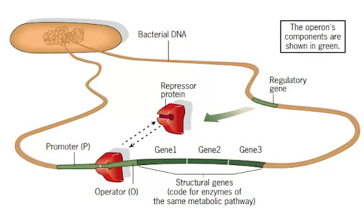
- The lac operon has lacZ, lacY, and lacA genes that code for galactosidase, galactose permease, and thiogalactoside transacetylase, respectively. These genes are preceded by an operator site (Olac) and a promoter (Plac).
- RNA polymerase copies the operon into a single polycistronic mRNA. This mRNA is then translated to make all three enzymes.
- When lactose is present, the background level of -galactosidase changes some lactose into allolactose. Allolactose then acts as an inducer and turns on transcription of the lac operon.
- IPTG can also be used to start a process.
- The lac repressor protein, which is made by the lacI gene, controls how much of the operon is copied.
- Lactose can be broken down by E. coli bacteria, but it’s not their favourite food. If glucose is available, they would use that much more. Lactose is harder to break down than glucose because it needs more steps and energy. But if lactose is the only sugar around, E. coli will use it right away as a source of energy.
- In order for the bacteria to use lactose, they must express the lac operon genes. These genes code for key enzymes that help the bacteria take in lactose and use it. For E. coli to be as efficient as possible, the lac operon should only be expressed when two conditions are met:
- Lactose is available, and
- Glucose is not available
- How are lactose and glucose levels measured, and how do changes in levels affect lac operon transcription? There are two regulatory proteins:
- One, the lac repressor, acts as a lactose sensor.
- The other, catabolite activator protein (CAP), acts as a glucose sensor.
- These proteins bind to the lac operon’s DNA and regulate its transcription in response to lactose and glucose concentrations.
Figure Schematic of the lac operonStructure of the lac operon
The components of a lac operon are Structural genes and
Regulatory DNA sequences.
Structural genes lac Operon
Structure of the lac operon
- The lac operon includes the lacZ, lacY, and lacA genes. These genes are controlled by a single promoter and transcribed as a single mRNA. Lac operon genes code for proteins that help the cell utilise lactose.
- lacZ: lacZ encodes the intracellular enzyme -galactosidase (LacZ), which hydrolyzes lactose into glucose and galactose.
- lacY: lacY encodes Beta-galactoside permease (LacY), a transmembrane symporter that uses a proton gradient in the same direction to pump -galactosides including lactose into the cell. Permease increases the cell’s permeability to -galactosides.
- lacA: lacA codes for the enzyme -galactoside transacetylase (LacA), which transfers an acetyl group from acetyl-CoA to thiogalactoside.
Regulatory DNA sequences
- The lac operon has several regulatory DNA sequences in addition to its three genes. These are sections of DNA to which particular regulatory proteins can bind, hence regulating transcription of the operon.
- Promoter: The promoter is the binding region for the enzyme that performs transcription, RNA polymerase.
- Operator: Lac repressor protein binds to the operator, a negative regulatory site. When the lac repressor is attached and the operator overlaps the promoter, RNA polymerase cannot connect to the promoter and initiate transcription.
- CAP: Catabolite activator protein binds to the CAP binding site, which is a positive regulatory site (CAP). When CAP is attached to this location, it facilitates the binding of RNA polymerase to the promoter, hence promoting transcription.
Llac repressor
- The lac repressor is a protein that suppresses (represses) lac operon transcription. This is accomplished by binding to the operator, which partially overlaps the promoter.
- When bound, lac repressor impedes RNA polymerase and prevents transcription of the operon.
- The gene lacI, which encodes the lac repressor, is under the control of its own promoter.
- The lacI gene is located in close proximity to the lac operon, however it is not a component of the operon and is expressed independently. lacI is continuously transcribed, therefore its protein product, lac repressor, is consistently present.
- When lactose is unavailable, the lac repressor binds strongly to the operator, inhibiting RNA polymerase transcription.
- In the presence of lactose, the lac repressor loses its capacity to bind DNA. It detaches itself from the operator, allowing RNA polymerase to transcribe the operon.
- This alteration in the lac repressor is caused by the lactose isomer allolactose.
- When lactose is present, certain molecules within the cell will be transformed to allolactose. Allolactose binds to lac repressor and causes it to shift form, preventing it from binding DNA.
- Allolactose is an example of an inducer, a tiny chemical that stimulates gene or operon expression.
- The lac operon is an inducible operon because it is normally turned off (repressed), but can be activated in the presence of allolactose.
LacI Function – Mechanism
- In the absence of lactose, LacI inhibits the synthesis of mRNA encoding proteins expressed by the lac operon.
- Although transcription is not eliminated entirely, lacZYA mRNA is transcribed at extremely low quantities. LacI protein binds with high specificity and affinity to the lac operator DNA sequence to perform this activity.
- Consequently, transcription is blocked by a variety of methods. Since the lac operator (LacO) overlaps the promoter, binding of LacI competes directly with RNA polymerase for binding to this region.
- LacI can also inhibit transcription start and/or mRNA elongation to effectively repress transcription.
- When lactose is available as a carbon source, lactose is transported into the bacterium by low quantities of the metabolic protein LacY.
- Next, residual LacZ converts lactose into glucose and galactose, which provides the bacterium with energy.
- Notably, this catalytic mechanism also produces low quantities of allolactose by rearranging the b-1,4 connection between glucose and galactose to a b-1,6 linkage.
- Allolactose binds to LacI and induces a conformational shift in the repressor protein, causing the operator DNA sequence to be released (induction).
- Thus, RNA polymerase is liberated to produce multiple copies of mRNA encoding lac metabolic proteins.
- Utilizing an environmental opportunity, these metabolic proteins, when translated into proteins, enable the bacterium to transport and digest huge volumes of lactose as its carbon energy source.
- Jacob and Monod discovered that a range of non-natural galactoside sugars (such as IPTG) can stimulate LacI and ease transcription inhibition of lacZYA as a consequence of their research. This discovery has had a significant impact on the biotechnology applications of this regulatory system.
(a) The lac repressor is active and binds the operator when allolactose is not present. Repressor binding to the operator inhibits transcription. (b) When lactose is available, some of it is converted to allolactose by β-galactosidase. When sufficient amounts of allolactose are present, it binds and inactivates the lac repressor. The repressor leaves the operator and RNA polymerase is free to initiate transcription.
Structural Characteristics of LacI
- The atomic-level process by which the LacI protein executes its tasks has been illuminated by LacI’s structural specifics
- LacI is a multimeric, multidomain protein, a configuration that is now recognised as being typical of genetic regulatory proteins.
- Four identical polypeptide chains comprise LacI. Each polypeptide folds into different functional areas, and four monomers combine to produce tetrameric LacI.
- Folded protein domains can be separated from the intact protein while retaining functions that are remarkably similar to those of the full protein.
- Proteolysis can be used to isolate the N-terminal domain of a LacI monomer, with or without portions of the hinge-helix sequence.
- An isolated N-terminal domain can bind with low affinity to one-half of LacO DNA. This protein fragment binds to full-length LacO with particularly high affinity if two of these domains and the hinge-helix sequences are connected by a disulfide bond (but is not inducible by galactoside sugars). (Note that the LacO site is strongly but imperfectly symmetric in sequence, allowing for the presence of two identical DNA-binding domains.)
- Only binding with high affinity by LacI inhibits mRNA transcription. Consequently, the LacI dimer is the functional unit, and a LacI tetramer has two DNA-binding sites.
- Each N-terminal domain of the LacI–LacO complex connects with DNA bases in the major groove, while the hinge helices insert into the minor groove to induce a DNA bend.
- LacI’s core domain is the second result of proteolysis. This segment is isolated as a tetramer, each monomer containing two subdomains that flank the sugar-binding site.
- Consequently, each monomer can bind an inducer molecule, resulting in four sugar-binding sites per tetramer. The N- and C-subdomains of the core domain monomer are coupled by numerous strands and cannot be dissociated by proteolysis.
- When the inducer-binding site is occupied in full-length LacI, the DNA-binding domain undergoes structural modifications in the N-subdomain.
- Because of these changes, the operator-specific binding affinity decreases, and the repressor protein that is bound to the inducer separates from the operator DNA site and moves to other parts of the genomic DNA.
- The C-terminal tetramerization domain of LacI joins two dimers by means of an antiparallel fourhelix bundle. When this domain is deleted, the resultant LacI dimer is still capable of repressing transcription.
Catabolite activator protein (CAP)
- Catabolite activator protein, CAP (also known as cAMP receptor protein, CRP) is essential for high levels of lac operon transcription.
- It forms a CRP–cAMP complex in association with 3’5′ cyclic AMP.
- CRP–cAMP binds to the lac promoter and enhances RNA polymerase binding, hence driving transcription of the lac operon.
- When glucose is available, intracellular cAMP levels decrease, CRP cannot bind to the lac promoter on its own, and the lac operon is only faintly transcribed.
- In the absence of glucose, intracellular cAMP levels increase, the CRP–cAMP complex is formed, and transcription of the lac operon is stimulated, allowing lactose to be used as an alternative carbon source.
- When lactose is present, lac repressor loses its capacity to bind to DNA. This allows RNA polymerase to connect to the lac operon’s promoter and initiate transcription.
- As it turns out, RNA polymerase alone does not bind the lac operon promoter particularly effectively. It might produce a few transcripts, but without the assistance of catabolite activator protein, it won’t accomplish much more (CAP).
- CAP attaches to a stretch of DNA immediately before the lac operon promoter and facilitates the attachment of RNA polymerase to the promoter, hence promoting high levels of transcription.
- CAP, like lac repressor, is encoded by a regulatory gene on the chromosome of bacteria. The CAP gene is not part of (or even close to) the lac operon.
- Constant or constitutive expression of the CAP gene. This indicates that CAP protein is constantly present in the cell to bind cAMP and “report” glucose levels to lac operon and other target genes and operons.
- CAP is not constantly active (able to bind DNA). Instead, it is controlled by cyclic AMP, a tiny molecule (cAMP). E. coli produces cAMP as a “hunger signal” when glucose levels are low.
- cAMP binds to CAP, changing its structure so that it can bind DNA and stimulate transcription. CAP cannot bind DNA and is inactive without cAMP.
- The act of transporting glucose into the cell decreases the creation of cAMP. Thus, when an abundance of glucose enters the cell, cAMP production is limited.
- If little or no glucose is available for transfer into the cell, however, cAMP synthesis is no longer blocked and cAMP levels increase. As a result: High glucose→Low cAMP; Low glucose→High cAMP
- CAP is only active at low glucose levels (cAMP levels are high). Thus, lac operon transcription is only possible at high levels in the absence of glucose. This technique ensures that bacteria activate the lac operon and begin utilising lactose only after depleting their preferred energy source (glucose).
Catabolite Repressor Protein
- Lactose is only energetically advantageous to bacteria when glucose is absent.
- Consequently, the lac operon contains two regulatory switches, one for each sugar. CRP is a protein that is utilised to assess glucose levels.
- This monitoring method is indirect and detects concentrations of the signalling chemical cAMP.
- cAMP is produced by a protein called adenylate cyclase, which is directly regulated (inhibited) by high glucose levels.
- Thus, glucose and cAMP levels are inversely connected. As glucose concentrations decline, cAMP concentrations rise, and cAMP binds to CRP. The CRP–cAMP complex has a strong affinity for target regions in the bacterial genome.
- One of the cAMP–CRP DNA target sequences is located within the promoter for lac metabolic proteins, upstream of the lac operator region.
- Note that cAMP binding increases CRP DNA binding, but sugar binding lowers DNA binding for LacI.
- DNA binding by the cAMP–CRP complex stimulates RNA polymerase transcription of lacZYA mRNA, whereas LacI binding inhibits this process.
- As a result, lactose metabolism proteins are produced at high levels only when ambient glucose levels are low and lactose levels are high.
CRP Structural Characteristics
- CRP, like LacI, is multimeric and composed of several domains.
- In this instance, the CRP is dimeric, with each monomer containing two domains.
- The N-terminal domain supports dimer formation and binds at least one cAMP molecule (two per dimer).
- In the absence of cAMP, the two monomeric DNA-binding domains are not adequately aligned for sequence-specific DNA recognition.
- In the presence of cAMP, the structure reorganises to place the two DNA-binding helix–turn–helix configurations in the correct orientation for efficient DNA binding.
- CRP–cAMP interaction also induces a DNA DNA-binding site bend, similar to LacI.
- Each monomer only attaches to one-half of the DNA-binding site, which is symmetrical along the sequence’s centre.
- Indeed, the symmetry observed in lac operon regulatory sites is a frequent characteristic of DNA regulatory binding sites, correlating with the multimeric nature of these proteins.
Multiple Sites/Multiple Targets – DNA Looping
- In addition to the primary operator, LacO, the lac operon sequence contains two ‘pseudo-operator’ sequences that contribute to repression.
- The DNA sequences of the pseudo-operators are quite close, but not identical, to those of LacO, and they are weakly bound by LacI.
- The presence of two DNA-binding sites in the LacI protein tetramer revealed a way by which pseudo-operators could augment repression – a single LacI protein tetramer could bind two distinct operators and produce a DNA structure with a loop.
- Multiple laboratories have produced experimental evidence for DNA looping. Recent research suggests that the angle between two dimers must widen for loop formation to take place.
- These highly stable looping structures account for the substantial lacZYA expression suppression reported in bacterial cells.
- Indeed, DNA having numerous operator sequences and possessing the E. coli-typical supercoiling density has a complicated half-life greater than 2 days.
- However, even these looped complexes respond quickly (in less than 30 s) to the presence of inducer sugars, allowing for rapid adaptation to a temporary external lactose source.
Common Errors in Understanding the lac Operon
A number of inaccuracies are regularly observed in
presentations of the lac operon
1st Error
- The most prevalent and widely disseminated misconception is that lactose is the inducer that relieves LacI suppression.
- According to this article, the molecule that functions as an inducer in vivo is allolactose, a lactose derivative produced by b-galactosidase as a byproduct of lactose hydrolysis to glucose and galactose.
- This ensures that the inducer is not created unless the metabolic capability to consume lactose is present in the cell.
2nd Error
- The second prevalent misconception is that lacZYA mRNA is never generated without lactose (and hence the inducer allolactose).
- Even in the absence of an inducer, typical dissociation events of LacI from LacO permit a little amount of read-through by RNA polymerase, resulting in extremely low levels of lacZYA mRNA production.
- This transcription ensures tiny amounts of lactose permease and b-galactosidase proteins are accessible to transport lactose and produce allolactose for induction.
- This leaky transcription can be a concern when the LacI/LacO system is employed to control the production of recombinant proteins: if the recombinant protein is toxic to E. coli, even minute amounts can be lethal.
- Thus, LacI/LacO regulation has been linked with other control systems (such as bacteriophage T7 polymerase) to impose a high level of repression until bacterial colonies reach large densities, and then to promote protein production.
3rd Error
- The third misconception is the notion that induced LacI cannot bind to the LacO operator (when bound to allolactose or gratuitous inducers).
- In reality, LacI inducer can bind to LacO, albeit with significantly lower strength.
- With this reduction in binding affinity, the LacI-inducer complex is unable to distinguish LacO from the rest of the DNA in the bacterial genome. The repressor protein is outcompeted by this surplus of nonspecific DNA, allowing RNA polymerase to transcribe lacZYA. This is because the genome is significantly larger than LacO, which represents a very, very small portion of the E. coli DNA sequence.
Conditions required to turn on lac operon
- The lac operon will be highly expressed if two conditions are met:
- Glucose must be absent: When glucose is absent, cAMP binds to CAP, allowing CAP to bind DNA. Bound CAP aids in the attachment of RNA polymerase to the lac operon promoter.
- Lactose must be available: If lactose is accessible, the operator will release the lac repressor (by binding of allolactose). This permits RNA polymerase to progress along the DNA and transcribe the operon.
Together, the engagement of the activator and the release
of the repressor allow RNA polymerase to bind tightly to the promoter and clear
the way for transcription. They result in robust transcription of the lac
operon and development of lactose-utilizing enzymes.
Lac operon Mechanism – In different Condition
Now that we’ve seen all the moving pieces of the lac operon,
let’s put what we’ve learned to use by seeing how the operon responds to
various situations (presence or absence of glucose and lactose).
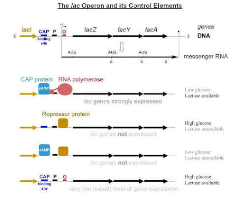
Glucose present, lactose absent: There is no
transcription of the lac operon. Because the lac repressor remains attached to
the operator and blocks RNA polymerase transcription, this is the case. Also,
because glucose levels are high and cAMP levels are low, CAP is inactive and
cannot bind DNA.
Glucose present, lactose present: There is low-level
transcription of the lac operon. Lac repressor is released from the operator
due to the presence of the inducer (allolactose). cAMP levels are however low
due to the presence of glucose. Thus, CAP remains inactive and is unable to
bind to DNA, limiting transcription to a low, leaky level.
Glucose absent, lactose absent: There is no
transcription of the lac operon. Since glucose levels are low and cAMP levels
are high, CAP is active and will bind to DNA. Due to the absence of allolactose,
the lac repressor will also bind to the operator, functioning as a blockage to
RNA polymerase and blocking transcription.
Glucose absent, lactose present: There is robust
transcription of the lac operon. Lac repressor is released from the operator
due to the presence of the inducer (allolactose). Due to the absence of
glucose, cAMP levels are up, therefore CAP is active and attached to DNA. CAP
facilitates the binding of RNA polymerase to the promoter, allowing for high
amounts of transcription.
Summary of lac operon responses
|
Glucose |
CAP binds |
Lactose |
Repressor binds |
Transcription |
|
+ |
– |
– |
+ |
No |
|
+ |
– |
+ |
– |
Some |
|
– |
+ |
– |
+ |
No |
|
– |
+ |
+ |
– |
Yes |
Lactose analogs
Several lactose derivatives or analogues that are suitable
for work with the lac operon have been described. The majority of these
substances are substituted galactosides in which the glucose moiety of lactose
has been replaced by another chemical group.
sopropyl-β-D-thiogalactopyranoside (IPTG)
- Isopropyl—D-thiogalactopyranoside (IPTG) is commonly employed as an inducer of the lac operon for physiological work.
- IPTG binds to repressor and renders it inactive, however it is not a substrate for -galactosidase.
- IPTG’s inability to be digested by E. coli is an advantage for its use in in vivo research.
- The rate of expression of lac p/o-controlled genes is not a variable in the experiment, nor is its concentration.
- In P. fluorescens, IPTG uptake is dependent on lactose permease activity, whereas in E. coli, this is not the case.
Phenyl-β-D-galactose (phenyl-Gal)
- Phenyl—D-galactose (phenyl-Gal) is a substrate for -galactosidase, however it is not an inducer because it does not deactivate repressor.
- Since wild-type cells produce so little -galactosidase, they cannot use phenyl-Gal as a carbon and energy source and so cannot grow on it. On phenyl-Gal, mutants devoid of repressor are able to grow.
- Therefore, minimum media containing only phenyl-Gal as a carbon and energy source is selective for repressor and operator mutants.
- When plating 108 cells of a wild-type strain on agar plates with phenyl-Gal, the rare colonies that develop are primarily spontaneous mutations that impact the repressor.
- The target size affects the relative distribution of repressor and operator mutations. Given that the lacI gene expressing repressor is approximately fifty times larger than the operator gene, repressor mutants prevail in the selection.
Thiomethyl galactoside [TMG]
- Another lactose homolog is thiomethyl galactoside (TMG).
- These suppress lacI repressor activity. At low inducer concentrations, the lactose permease allows both TMG and IPTG to enter the cell.
- At high inducer concentrations, however, both analogues can independently reach the cell. TMG can inhibit cell proliferation at high extracellular doses.
Other compounds for colorful indicators of
β-galactosidase activity
- Other substances function as coloured -galactosidase activity indicators.
- ONPG: ONPG is typically utilised as a substrate for in vitro assays of -galactosidase since it is cleaved into the highly yellow chemical ortho nitrophenol and galactose.
- X-gal (5-bromo-4-chloro-3-indolyl—D-galactoside) is an artificial substrate for B-galactosidase whose cleavage results in galactose and 4-Cl,3-Br indigo, providing a rich blue colour in colonies that manufacture -galactosidase.
Allolactose
- Allolactose is the inducer of the lac operon and is an isomer of lactose.
- Allolactose consists of galactose-β(1→6)-glucose, whereas lactose consists of galactose-β(1→4)-glucose.
- In an alternate process to the hydrolytic one, lactose is transformed to allolactose by -galactosidase.
- A physiological experiment demonstrating the role of LacZ in the generation of the “real” inducer in E. coli cells is the observation that a lacZ null mutant may still produce LacY permease when cultivated with IPTG, a non-hydrolyzable analogue of allolactose, but not with lactose.
- The conversion of lactose to allolactose, catalysed by -galactosidase, is necessary for the production of the inducer within the cell.
Lac operon Important Notes
Lac operon comprises metabolically relevant genes.
- The genes are exclusively expressed in the presence of lactose and the absence of glucose.
- In reaction to glucose and lactose levels, the catabolite activator protein and lac repressor operon is turned on and off.
- The lac repressor inhibits the operon’s transcription. In the presence of lactose, its repressor function ceases.
- When glucose levels are low, catabolite activator protein initiates the transcription of the operon.
Regulation of Lac Operon – Positive and Negative
- The lac operon is an excellent example of negative control (negative regulation) of gene expression, as binding repressor inhibits transcription of structural genes.
- Positive gene expression control or regulation occurs when the regulatory protein attaches to DNA and enhances the transcription rate.
- This regulating protein is referred to as an activator. The lac operon regulator CAP/CRP is an excellent example of an activator.
- Consequently, the lac operon is sensitive to both negative and positive regulation.
Lac Operon Regulation by cyclic AMP
- The explanation for diauxie required the identification of additional mutations affecting the lac genes, in addition to those accounted for by the traditional hypothesis.
- Two more genes, cya and crp, were subsequently found that mapped far from lac and that, when mutated, result in a reduction in expression in the presence of IPTG and in strains of the bacteria without the repressor or operator.
- The discovery of cAMP in E. coli led to the revelation that mutants deficient in the cya gene, but not the crp gene, might have their activity restored by adding cAMP to the media.
- cAMP is produced by adenylate cyclase, which is encoded by the cya gene. In a cya mutant, the absence of cAMP reduces lacZYA gene expression by more than tenfold compared to normal.
- Adding cAMP corrects the reduced Lac expression observed in cya mutants. Catabolite activator protein (CAP) or cAMP receptor protein is encoded by the second gene, crp (CRP).
- However, lactose metabolism enzymes are produced in small amounts in the presence of both glucose and lactose (sometimes referred to as leaky expression) because the LacI repressor rapidly associates/dissociates from the DNA as opposed to tightly binding to it, allowing RNAP to bind and transcribe mRNAs of lacZYA.
- Leaky expression is required so that some lactose can be metabolised after the glucose source has been depleted, but before lac expression is fully triggered.
- The time required to manufacture sufficient numbers of lactose-metabolizing enzymes is reflected in the interval between growth phases.
- First, the CAP regulatory protein must assemble on the lac promoter, hence increasing lac mRNA output.
- Significantly more LacZ (-galactosidase, for lactose metabolism) and LacY (-galactosidase, for lactose metabolism) copies are produced when there are more lac mRNA copies (lactose permease to transport lactose into the cell).
- After a delay necessitated by the elevation of lactose-metabolizing enzymes, the bacteria undergo a new phase of fast cell division.
- Catabolite suppression presents two mysteries: first, how cAMP levels are connected to the presence of glucose, and second, why the cells bother.
- After lactose is cleaved, glucose and galactose are produced (easily converted to glucose). In terms of metabolism, lactose is an equivalent carbon and energy source to glucose.
- The quantity of cAMP is not connected to intracellular glucose concentration, but rather to the pace of glucose transport, which affects the activity of adenylate cyclase. Additionally, glucose transport inhibits lactose permease directly. As to why E. coli behaves in this manner, only speculation can be made.
- All intestinal bacteria digest glucose, indicating that they are frequently exposed to it. It may be advantageous for cells to regulate the lac operon in this manner due to a little differential in the efficiency of transport or metabolism between glucose and lactose.
What Do You Mean by Inducers and the Induction of Lac operon?
- Normally, E. coli cells produce relatively little of each of these three proteins, but when lactose is present, the amount of each enzyme increases dramatically and in a coordinated manner.
- Consequently, every enzyme is an inducible enzyme, and the process is known as induction.
- The few molecules of ß-galactosidase in the cell prior to induction convert lactose to allolactose, which subsequently activates the transcription of these three lac operon genes.
- Allolactose is therefore an inducer.
- Another lac operon inducer is isopropylthiogalactoside (IPTG). Unlike allolactose, this inducer is not metabolised by E. coli and is therefore solely effective for induction experiments.
Lac Operon when Inducers are not present
In the absence of an inducer such as allolactose or IPTG,
the lacI gene is transcribed and the resultant repressor protein binds to the
operator site of the lac operon, Olac, thereby inhibiting transcription of the
lacZ, lacY, and lacA genes.
Lac Operon when Inducers are present
Induction involves the binding of the inducer to the
repressor. This alters the structure of the repressor, reducing its affinity
for the lac operator site significantly. The lac repressor has now dissociated
from the operator site, permitting the RNA polymerase (already present on the
neighbouring promoter site) to initiate transcription of the lacZ, lacY, and
lacA genes. They are transcribed to form a single polycistronic mRNA, which is
subsequently translated into vast quantities of all three enzymes. The presence
of polycistronic mRNA guarantees that the concentrations of all three gene
products are coordinatedly regulated. If the inducer is eliminated, the lac
repressor attaches fast to the lac operator site and transcription is virtually
instantly stopped.
References
David Hames and Nigel Hooper (2005). Biochemistry. Third ed.
Taylor & Francis Group: New York.
Parija S.C. (2012). Textbook of Microbiology &
Immunology.(2 ed.). India: Elsevier India.
Lehninger Principles of Biochemistry: International Edition
by David L. Nelson (Author), Michael Cox
(Author)
Molecular Biology by Robert F. Weaver (Author)
Osbourn AE, Field B. Operons. Cell Mol Life Sci. 2009
Dec;66(23):3755-75. doi: 10.1007/s00018-009-0114-3. Epub 2009 Aug 7. PMID:
19662496; PMCID: PMC2776167.
Swint-Kruse, L., & Matthews, K. S. (2013). lac Operon.
Encyclopedia of Biological Chemistry, 694–700.
doi:10.1016/b978-0-12-378630-2.00249-8
https://www.khanacademy.org/science/ap-biology/gene-expression-and-regulation/regulation-of-gene-expression-and-cell-specialization/a/the-lac-operon


No comments